
Researchers reconnect the completely divided spinal cord of a rat thanks to graphene foams
The Materials Science Institute of Madrid (ICMM-CSIC), part of the Ministry of Science, Innovation and Universities (MICIU), has reconnected, in a rat, a completely sectioned spinal cord at the thoracic level thanks to a three-dimensional foam created with reduced graphene oxide. The work, just published in the journal Bioactive Materials, demonstrates the potential of this material for the treatment of spinal cord injuries, and opens new avenues of research towards the cure of paraplegic patients in different states of the disease.
When a spinal cord injury occurs, it is usually not a complete break, but the injury typically affects only a specific part, at one or more levels of the spinal cord. Nevertheless, this study aims to demonstrate that this material can enhance the reconnection of neural tissue even when the injury is complete. Conchi Serrano, a researcher at ICMM-CSIC and one of the main authors of the work, explains: "Our team had already demonstrated that these foams create a pro-repair environment in the rat spinal cord, but we wanted to extend this by increasing the size of the injury and changing the spinal level, and we have managed to replicate the results."
This group, in close collaboration with researchers from the National Hospital of Paraplegics in Toledo such as Juan Aguilar and Elisa López, prepared a foam (called a scaffold) made of reduced graphene oxide: "It undergoes a thermal treatment at 220°C to eliminate excess oxygen groups and increase chemical bonds between sheets, which gives us greater mechanical stability," explains Serrano, who has been working on this material for neural regeneration applications for over a decade.
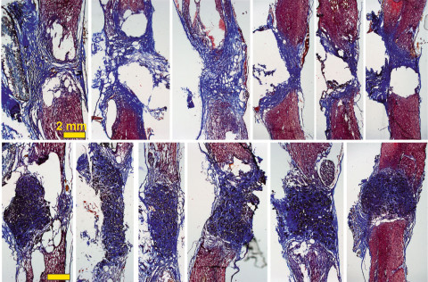
Thus, when the scaffold is placed in the spinal cord—in this case, in a rat model with the spinal cord completely sectioned at the thoracic level—"we see a large number of blood vessels, which are crucial for nourishing the new tissue, as well as neurites (the filaments that connect one neuron to another)." The researcher explains that they observe "how the neurons that have survived in the area around the injury extend their projections through the scaffold and invade it in its full 3D extent." All of this improves over time: the results are evident after 10 days of implantation, but they are much more promising after 4 months.
"Our reduced graphene oxide scaffolds promote the growth of more abundant and larger blood vessels, as well as more abundant, longer neurites that are more evenly distributed throughout the injury space," celebrates Serrano.
Additionally, the researchers carried out electrophysiological recordings. They observed the brain's response when stimulating the spinal cord below the damaged area, and the results are revealing: "We recorded a response in the brain, confirming not only that neural tissue is crossing through the scaffold, but also that it is reconnecting with the brain." Specifically, the response is seen in the reticular formation, a functionally significant area for motor function.
This work is part of the Piezo4Spine project, funded by the European Union through the Horizon Europe Pathfinder program, which aims to cure spinal cord injuries through nanotechnology. With this goal, nanomedicines are also being developed, which in the next phase of this work will be incorporated into the scaffold to further promote these promising regenerative findings.
Referencia:
Marta Zaforas, Esther Benayas, Raquel Madroñero-Mariscal, Ana Domínguez-Bajo, Elena Fernández-López, Yasmina Hernández-Martín, Ankor González-Mayorga, Elena Alonso-Calviño, Eduardo R. Hernández, Elisa López-Dolado, Juliana M. Rosa, Juan Aguilar*, María C. Serrano*. Graphene oxide scaffolds promote functional improvements mediated by scaffold-invading axons in thoracic transected rats. Bioactive Materials. DOI: https://doi.org/10.1016/j.bioactmat.2024.12.031
ICMM Comunicación - comunicacion@icmm.csic.es
Instituto de Ciencia de Materiales de Madrid (ICMM)
Sor Juana Ines de la Cruz, 3
Cantoblanco, 28049
Madrid, España
Telephone: (+34) 91 334 90 00
Email: @email
Communication Office: @email

Acknowledge the Severo Ochoa Centres of Excellence program through Grant CEX2024-001445-S/ financiado por MICIU/AEI / 10.13039/501100011033

Contacto | Accesibilidad | Aviso legal | Política de Cookies | Protección de datos