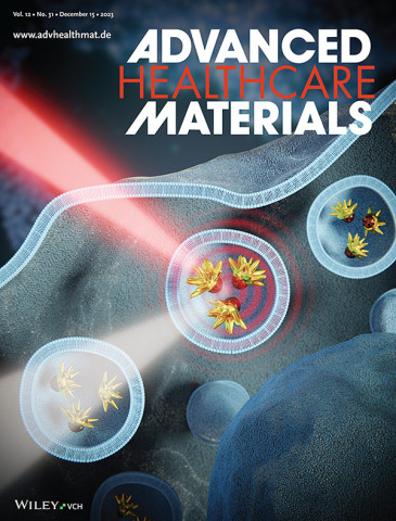
Researchers manage to monitor the local temperature of nanoparticles in tumor cells with X-rays

Scientists from the Materials Science Institute of Madrid (ICMM-CSIC) have developed a technique for directly measuring the temperature of nanoparticles inside human tumor cells using X-rays. This advancement could lead to the design of more precise and less toxic therapies for cancer treatment through hyperthermia, a technique that aims to cure tumors by raising their temperature.
Hyperthermia is often used alongside radiotherapy and chemotherapy in clinical cancer treatment. The use of heat-generating nanoparticles (in the range of one billionth of a meter) offers multiple advantages, although measuring temperature at this scale (known as nanothermometry) poses a significant challenge. This is where this work represents a milestone.
For nanothermometry, it is essential to measure and monitor the temperature of nanoparticles internalized in tumor cells and understand how they react to these temperatures. "The challenge is to obtain 'nano' thermometers that are sensitive and robust in the biological environment and operational over a wide temperature range," explains Ana Espinosa, a researcher at ICMM-CSIC and the lead author of the study, along with Álvaro Muñoz Noval from the Complutense University of Madrid (UCM).
Until now, very precise optical techniques based on the fluorescence of certain added particles have been used for these measurements. Still, this team has gone a step further by exploring a new X-ray technique that allows the direct measurement of temperature inside the cells being studied without the need for additional markers. On one hand, the nanoparticles receive heat through near-infrared light, while on the other hand, X-rays are used to observe the temperature increase and its effect on the cell.
"It is a direct measurement method: atoms vibrate and heat up, which is captured by X-rays without the need for another particle to reflect that change in temperature," indicates Espinosa, noting that, although gold and iron oxide nanoparticles were used in the study, they could be made of another material.
The key result of this study has been the observation of how, when applying heat under these conditions, nanoscale temperatures are higher than at macroscopic levels. This will be crucial for designing more precise and less toxic therapies in every aspect: the temperature applied to cells can be reduced, requiring less laser power, fewer nanoparticles will be needed, and the treatment can be shorter.
Furthermore, this nanometric thermometer can be universally applied, as it can be used in any process requiring temperature, such as in microelectronics, catalysis, etc. "It operates effectively over a wide range of temperatures, becoming a versatile tool for studying thermal fluctuations in general," argues the researcher.
Due to the high sensitivity required for the study, measurements were conducted at the SAMBA beamline at the SOLEIL synchrotron (Saint-Aubin, France). Still, the goal is to be able to perform it in the future with laboratory instrumentation. The work, published in the journal Advanced Healthcare Materials, used an in vitro 3D tumor model of glioblastoma (a type of brain cancer).
This study is a collaboration between researchers from various centers: IMDEA Nanociencia (Spain), BCMaterials (Spain), SOLEIL synchrotron (France), Curie Institute (France), Institute of Ceramics and Glass (ICV-CSIC, Spain), Institute of Materials Science (ICMM-CSIC, Spain), and Complutense University of Madrid (Spain).
Reference:
R. López-Méndez, J. Reguera, A. Fromain, E. Samy Abu Serea, E. Céspedes, F. Jose Teran, F. Zheng, A. Parente, M.A. García, E. Fonda, J. Camarero, C. Wilhelm, Á. Muñoz-Noval, A. Espinosa: X-Ray Nanothermometry of Nanoparticles in Tumor-Mimicking Tissues under Photothermia, Advanced Healthcare Materials (2023), DOI: 10.1002/adhm.202301863
ICMM Comunicación/ CSIC Comunicación - @email
Instituto de Ciencia de Materiales de Madrid (ICMM)
Sor Juana Ines de la Cruz, 3
Cantoblanco, 28049
Madrid, España
Telephone: (+34) 91 334 90 00
Email: @email
Communication Office: @email

Acknowledge the Severo Ochoa Centres of Excellence program through Grant CEX2024-001445-S/ financiado por MICIU/AEI / 10.13039/501100011033

Contacto | Accesibilidad | Aviso legal | Política de Cookies | Protección de datos
