We develop new dynamic AFM modes and methodologies to obtain high resolution images of crystalline surfaces, biomolecules and hydration layers in different liquids. The key point is to operate the instrument under very small forces (below 100 pN). For example, we have generated molecular resolution images of different biological samples in physiological environment. Bacteriorhodopsin crystals (Figure 1), GroEL molecules (Figure 2) and IgG antibodies were imaged in liquid environment with nanometer spatial resolution.
Lower lateral and vertical forces are required to measure single molecules like IgG or GroEL as compared to 2D crystals such as Bacteriorhodopsin.An example of the spatial resolution provided by advanced dynamic AFM we have imaged the formation of hydration layers on muscovite mica (Figure 4).
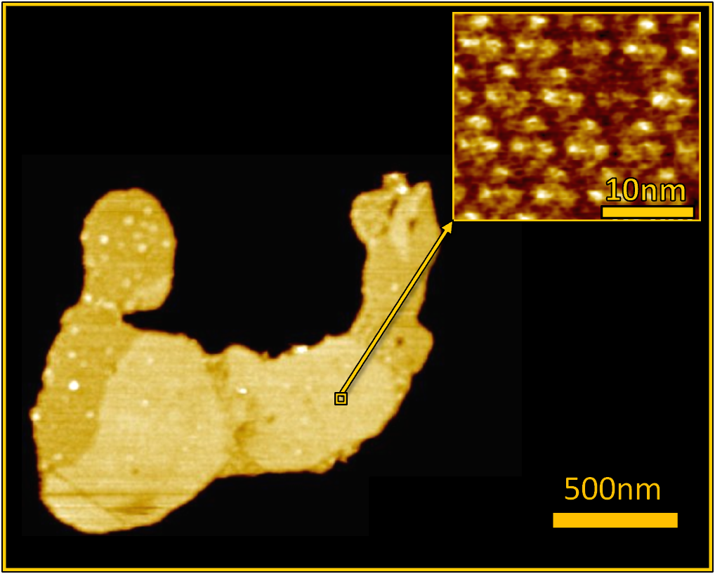
Figure 1: Zoom of a patch of Purple Membrane. Bacteriorhodopsins with trimeric structure form a two dimensional crystal.
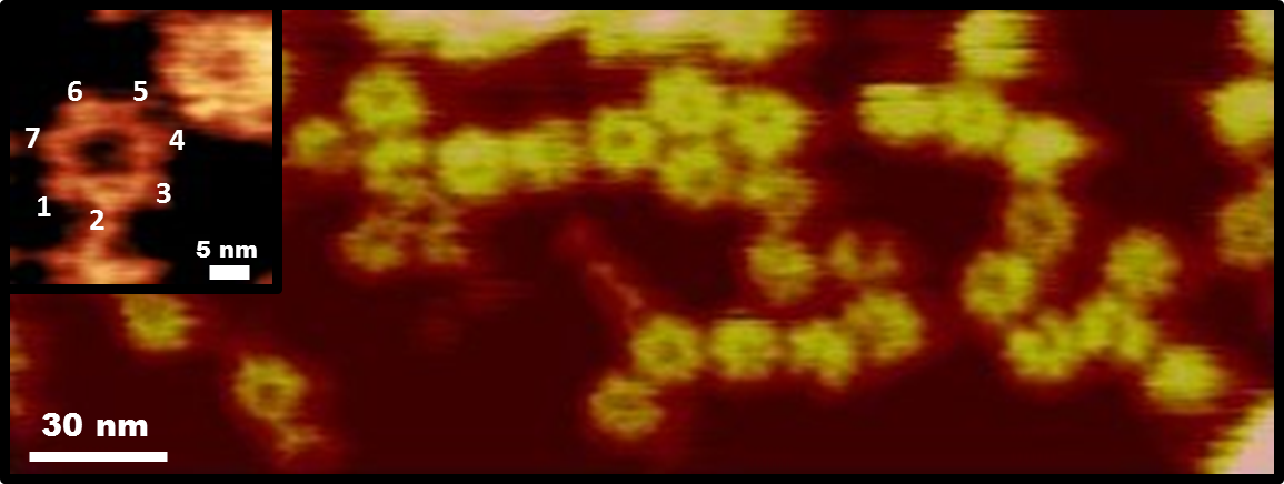
Figure 2: Image of a GroEL protein in buffer. In the inset, the heptameric ring structure is shown.
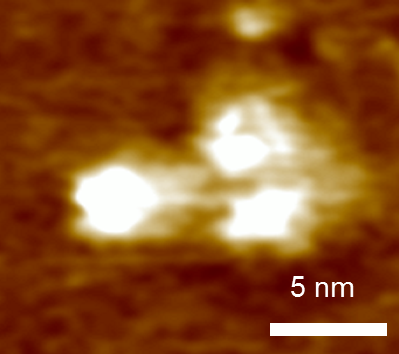
Figure 3: Single IgG antibody.
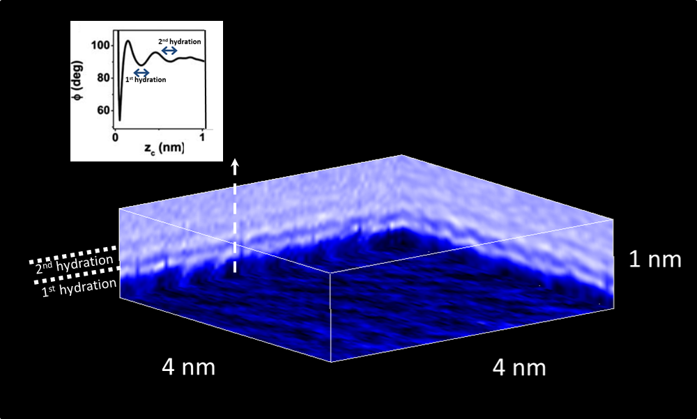
Figure 4: Three-dimensional image of muscovite mica in a KCl solution. The first and the second hydration layers are observed
Publications:
- Elena T. Herruzo, Hitoshi Asakawa, Takeshi Fukuma and Ricardo Garcia. Nanoscale 5, 2678-2685 (2013)
- Amir F. Payam, Jorge R. Ramos and Ricardo García. ACS Nano 6, 4663-4670 (2012)
- S. Patil, N. F. Martínez, J. R. Lozano and R. García. Journal of Molecular Recognition 20, 516-523 (2007)
- D. Martínez-Martín, E. T. Herruzo, C. Dietz, J. Gómez-Herrero, and R. García. Physical Review Letters 106, 198101 (2011)